VR developed by Albert Liu, Ahmet Emre, Nicholas Kotov, Syahidah Mohd Khairi, Trevor Teague and Theodore Hall
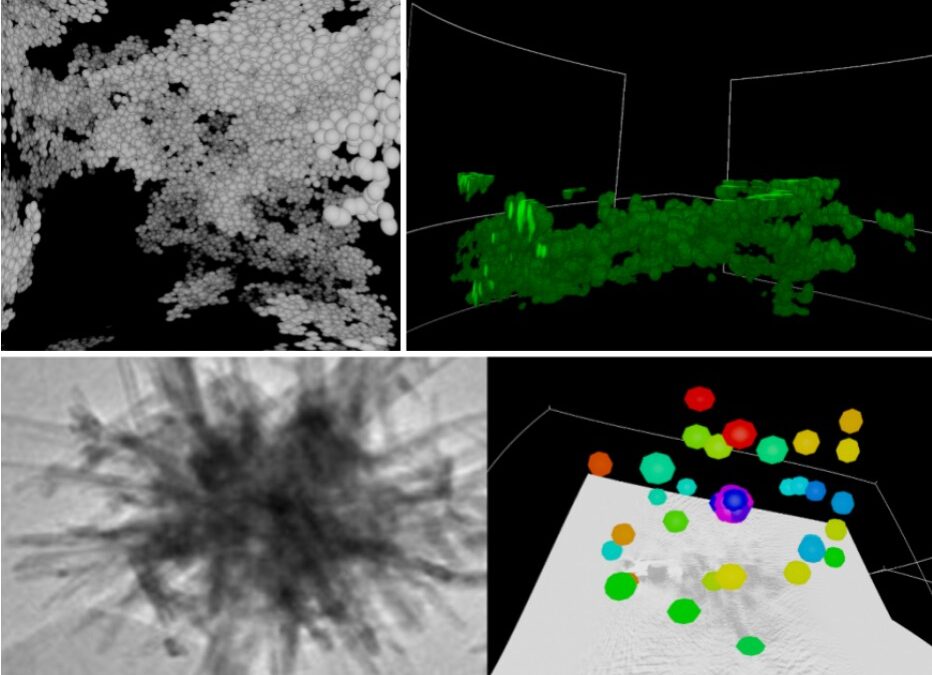
The second module focuses on the microscopy of small-scale systems, where the objective is to help students explore 3D imaging techniques (optical and electron microscopy) to visualize and characterize the particles and particle systems. There are 3 activities: (1) Percolation Simulation, (2) Colloidal Gel Confocal Microscopy, and (3) Hedgehog Particle Electron Microscopy.
 To paint on the scene in 3D, you can use the X and Y (left-hand) or A and B (right-hand) buttons. Press and hold Y or B to paint in 3D, and press X or A to clear the paint. You will use this feature to annotate the 3D scene in your lab write-up. The two controllers are symmetric. Press and hold the side trigger with your middle finger (either hand) and “drag” to use the virtual 3D Tape Measure. The feedback displays dX, dY, dZ, dL: the first three numbers are the changes in the X, Y, and Z coordinates; the last number is the length or distance measured.
Activity 1: Percolation of particles
This activity introduces a percolated particle network, where the structure spans the entire system. Users can explore a simulated network of tetrahedra and spheres through interactive fly-through visualization, allowing observation of connectivity and overall structure within the system.
Activity 2: Colloidal gel microscopy
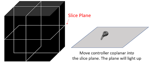
This activity presents a 3D confocal microscopy analysis of a colloidal gel system, enabling exploration through planar slicing and fly-through visualization of spherical particle networks. The image stacks are obtained using fluorescence microscopy, consisting of x–y scans at varying heights. By navigating and slicing the data in different directions, users can simulate 3D image construction from stacked 2D images and perform basic particle characterization, such as measuring particle size along different axes.
Activity 3: Hedgehog particle microscopy
This activity provides an exploration of the 3D microscopy of a hedgehog-shaped particle by slicing the image along multiple planes to observe its structure from different perspectives. Hedgehog particles are engineered to increase surface area and have applications in areas such as catalysis and energy storage. The imaging data are obtained using TEM (transmission electron microscopy) tomography. The activity also introduces image-based particle characterization, allowing users to annotate features such as the vertices of the hedgehog structure, demonstrating key analysis methods used with 3D microscopy data.
We also have a CAVE version of Colloidal Gel Confocal Microscopy for the students to collaborate with each other in understanding the 3D microscopy images.